| Cat # | Size | Price | Quantity | |
|---|---|---|---|---|
| 117401 | 25 µg | $150 | ||
| 117402 | 100 µg | $325 |
| Clone | ICASG1AB |
|---|---|
| Application | Flow Cytometry, ICC |
| Reactivity | Human |
| Format | Purified |
| Target Name | ASGR1, ASGPR1 |
| Isotype | Rabbit IgG |
| Antibody Type | Monoclonal |
| Regulatory Status | RUO |
| Formulation | Phosphate-buffered solution, pH 7.2, containing 0.09% sodium azide |
| Protein Concentration | 0.5 mg/mL |
| Storage&Handling | The antibody solution should be stored between 2°C and 8°C |
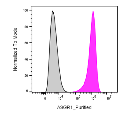
| Recommended Usage | For flow cytometric staining, it is recommended to use less than 0.2 µg of this reagent per 0.5-1.0 million cells in a 100 µL volume. Optimal reagent performance should be determined by titration for each specific application |
| See All Formats | Clone ICASG1AB |
Human ASGR1 (asialoglycoprotein receptor 1) is a type II transmembrane lectin primarily expressed on the surface of hepatocytes in the liver. It forms the major subunit of the asialoglycoprotein receptor (ASGPR), a receptor complex responsible for the rapid clearance of circulating glycoproteins that have lost their terminal sialic acid residues. By recognizing exposed galactose or N-acetylgalactosamine (GalNAc) residues, ASGR1 mediates receptor-dependent endocytosis and lysosomal degradation of these glycoproteins, thereby maintaining serum glycoprotein homeostasis and contributing to liver-specific clearance of endogenous and exogenous molecules.
Structurally, ASGR1 is a calcium-dependent C-type lectin receptor composed of a short cytoplasmic tail, a single transmembrane domain, a stalk region, and a C-terminal carbohydrate recognition domain (CRD). The receptor typically forms hetero-oligomeric complexes with ASGR2, which enhance ligand binding and internalization efficiency. Its principal ligands include desialylated glycoproteins bearing terminal galactose or GalNAc residues, and ligand binding requires calcium ions for high-affinity recognition.
ASGR1 has been implicated in lipid metabolism and cardiovascular disease. Naturally occurring loss-of-function variants in ASGR1 are associated with reduced levels of non-high-density lipoprotein cholesterol and a lower risk of coronary artery disease, suggesting that the receptor influences cholesterol homeostasis in addition to glycoprotein clearance. Altered ASGR1 expression has also been investigated in liver diseases, although its precise contribution to hepatic pathology remains under active study.
ASGR1 has become an important therapeutic target because its highly selective expression on hepatocytes enables liver-specific drug delivery. Triantennary GalNAc conjugates bind ASGR1 with high affinity and efficiently deliver small interfering RNAs, antisense oligonucleotides, and other nucleic acid therapeutics into hepatocytes. This targeting strategy has substantially improved the efficacy and safety of numerous liver-directed therapies and represents a major advance in precision medicine for metabolic, genetic, and infectious liver diseases.
Rabbit IgG, κ2 Isotype Control Antibody
Have a product or application question? Consult our FAQs or contact us.