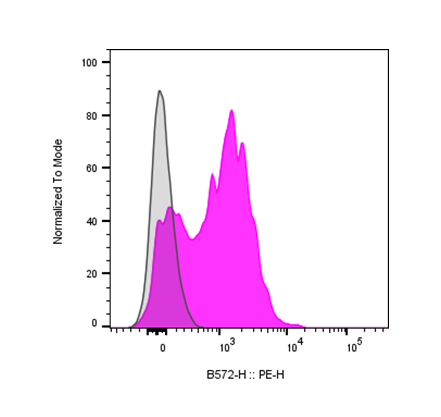
| Cat # | Size | Price | Quantity | |
|---|---|---|---|---|
| 110201 | 100 ug | $140 | ||
| 110202 | 500 ug | $320 |
| Clone | 184AM1 |
|---|---|
| Application | Flow Cytometry |
| Reactivity | Human |
| Format | Purified |
| Target Name | CD184, CXCR4 |
| Isotype | Mouse IgG1 |
| Antibody Type | Monoclonal |
| Regulatory Status | RUO |
| Formulation | Phosphate-buffered solution, pH 7.2, containing 0.09% sodium azide |
| Protein Concentration | 0.5 mg/mL |
| Storage&Handling | The antibody solution should be stored between 2°C and 8°C |
| Recommended Usage | For flow cytometric staining, it is recommended to use less than 0.25 µg of this reagent per 0.5-1.0 million cells in a 100 µL volume. Optimal reagent performance should be determined by titration for each specific application |
| RRID | AB_3738938 |
| Research Areas | Chemokine receptors, HIV, T cells, B cells, Monocytes, Macrophages, Neutrophils, Eosinophils, NK cells, Hematopoietic Stem cells, Endothelial cells, Astrocytes, Neurons, Chemotaxis, Development, Infectious Diseases, Cancer Marker |
| See All Formats | Clone 184AM1 |
CD184, also known as CXCR4 (C-X-C chemokine receptor type 4), is a G protein-coupled receptor (GPCR) that plays a central role in cell migration, immune regulation, and development. It is expressed on a wide variety of cells, including hematopoietic stem and progenitor cells, lymphocytes, endothelial cells, and many types of cancer cells. CXCR4 primarily functions as a chemokine receptor that guides cell trafficking and positioning in response to chemotactic gradients, making it essential in immune surveillance, hematopoiesis, and organogenesis.
Structurally, CD184 is a typical seven-transmembrane GPCR composed of 352 amino acids with an extracellular N-terminal domain, seven hydrophobic transmembrane helices, three extracellular and intracellular loops, and a cytoplasmic C-terminal tail. The extracellular regions are responsible for ligand binding, while the intracellular domains interact with G proteins and β-arrestins to initiate downstream signaling cascades. The receptor is heavily glycosylated at the N-terminus, which contributes to ligand specificity and receptor stability. The cognate ligand for CD184 is CXCL12, also known as stromal cell–derived factor 1 (SDF-1). Binding of CXCL12 to CXCR4 triggers activation of multiple signaling pathways, including PI3K/AKT, MAPK/ERK, and calcium mobilization cascades, which collectively regulate migration, survival, and proliferation. The CXCL12-CXCR4 axis is critical for the homing of hematopoietic stem cells to the bone marrow and the migration of immune cells during inflammation and tissue repair.
CD184 has been implicated in numerous diseases, ranging from cancer to infectious and autoimmune disorders. In oncology, CXCR4 is frequently overexpressed in solid and hematologic malignancies, where it drives tumor growth, angiogenesis, metastasis, and resistance to therapy. The receptor also serves as a co-receptor for HIV-1 entry into T cells, enabling viral infection and progression to AIDS. Dysregulation of the CXCL12-CXCR4 axis is additionally associated with inflammatory diseases, such as rheumatoid arthritis and multiple sclerosis, due to aberrant immune cell recruitment.
Therapeutically, CXCR4 represents a major target in both cancer and regenerative medicine. Antagonists such as plerixafor (AMD3100) are used clinically to mobilize hematopoietic stem cells for transplantation. In cancer, CXCR4 inhibitors and monoclonal antibodies are under development to block metastasis and improve immunotherapy efficacy. Modulating this pathway continues to hold significant potential for controlling immune responses, enhancing stem cell therapies, and limiting disease progression across multiple clinical contexts.
Mouse IgG1 Isotype Control Antibody
Anti-Human CD184 (CXCR4) Antibody TDS
Anti-Human CD184 (CXCR4) Antibody, Clone 184AR1
PE Anti-Human CD184 (CXCR4) Antibody, Clone 184AR1
Have a product or application question? Consult our FAQs or contact us.