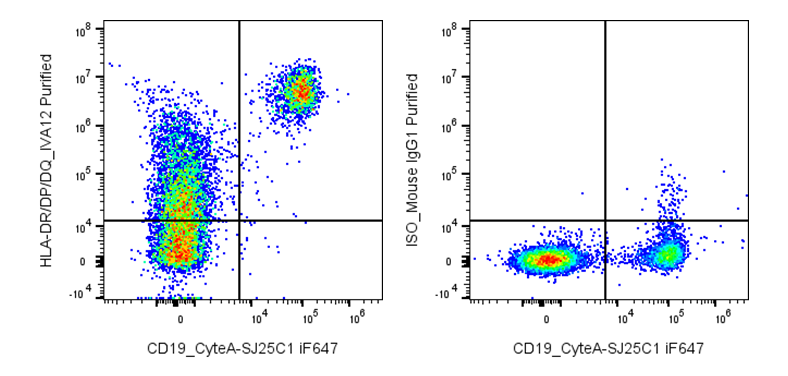
| Cat # | Size | Price | Quantity | |
|---|---|---|---|---|
| 118901 | 25 µg | $35 | ||
| 118902 | 100 µg | $95 |
| Clone | IVA12 |
|---|---|
| Application | Flow Cytometry |
| Reactivity | Human |
| Format | Purified |
| Target Name | HLA-DR, HLA-DP, and HLA-DQ, MHC class II, pan HLA-II, pan MHC-II |
| Isotype | Mouse IgG1 |
| Antibody Type | Monoclonal |
| Regulatory Status | RUO |
| Formulation | Phosphate-buffered solution, pH 7.2, containing 0.09% sodium azide |
| Protein Concentration | 0.5 mg/mL |
| Storage&Handling | The antibody solution should be stored between 2°C and 8°C |
| See All Formats | Clone IVA12 |
Human HLA-DR, HLA-DP, and HLA-DQ are the three major classical human leukocyte antigen (HLA) class II molecules encoded within the major histocompatibility complex (MHC) on chromosome 6. These cell-surface glycoproteins are primarily expressed on professional antigen-presenting cells, including dendritic cells, macrophages, and B lymphocytes. Their principal function is to present peptides derived from extracellular proteins to CD4+ T helper cells, initiating and regulating adaptive immune responses. This process is essential for host defense against pathogens and for the development of immune memory.
Each HLA class II molecule consists of a polymorphic α (alpha) chain and β (beta) chain that together form a peptide-binding groove capable of accommodating peptides approximately 13–25 amino acids in length. The groove binds peptides generated in endosomal and lysosomal compartments following antigen processing. Peptide loading is facilitated by accessory molecules such as HLA-DM after removal of the invariant chain-derived CLIP peptide. The extensive genetic polymorphism of HLA-DR, HLA-DP, and HLA-DQ enables presentation of a broad repertoire of peptide ligands, including self and foreign antigens.
Specific HLA class II alleles are strongly associated with autoimmune diseases. For example, HLA-DR and HLA-DQ variants contribute to susceptibility to type 1 diabetes, rheumatoid arthritis, celiac disease, multiple sclerosis, and systemic lupus erythematosus. Certain HLA-DP alleles have also been linked to chronic hepatitis B outcomes and other immune-mediated disorders. These molecules are important in transplantation because mismatched HLA alleles increase the risk of graft rejection.
Therapeutically, HLA-DR, HLA-DP, and HLA-DQ are used in tissue matching for organ and stem cell transplantation, disease risk assessment, vaccine development, and immunotherapy. Their ability to present antigenic peptides also makes them attractive targets for antigen-specific tolerance strategies and personalized immunotherapies designed to modulate pathogenic T-cell responses while preserving protective immunity.
Mouse IgG1 Isotype Control Antibody
Anti-Human HLA-DR/DP/DQ Antibody TDS
Have a product or application question? Consult our FAQs or contact us.