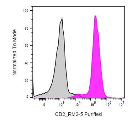
| Cat # | Size | Price | Quantity | |
|---|---|---|---|---|
| 207301 | 25 µg | $30 | ||
| 207302 | 100 µg | $60 |
| Clone | RM2-5 |
|---|---|
| Application | Flow Cytometry, IP, Costimulation, Block |
| Reactivity | Mouse |
| Format | Purified |
| Target Name | CD2, LFA-2, T11, Ly-37, SRBC-R |
| Isotype | Rat IgG2b |
| Antibody Type | Monoclonal |
| Regulatory Status | RUO |
| Formulation | Phosphate-buffered solution, pH 7.2, containing 0.09% sodium azide |
| Protein Concentration | 0.5 mg/mL |
| Storage & Handling | The antibody solution should be stored between 2°C and 8°C |
| Recommended Usage | For flow cytometric staining, it is recommended to use less than 0.2 µg of this reagent per 0.5-1.0 million cells in a 100 µL volume. Optimal reagent performance should be determined by titration for each specific application |
| See All Formats | Clone RM2-5 |
Mouse CD2 is a cell surface glycoprotein of the immunoglobulin superfamily expressed primarily on T lymphocytes, natural killer (NK) cells, and a subset of thymocytes. It functions as an adhesion and costimulatory receptor that promotes stable interactions between immune cells, facilitating antigen recognition, immune synapse formation, and lymphocyte activation. CD2 lowers the threshold for T-cell receptor (TCR) signaling by strengthening contact between T cells and antigen-presenting cells (APCs), thereby enhancing T-cell proliferation, cytokine production, and cytotoxic effector functions. In NK cells, CD2 similarly augments activation and target cell killing through cooperative signaling with other activating receptors.
Structurally, mouse CD2 is a type I transmembrane glycoprotein consisting of two extracellular immunoglobulin-like domains, a single transmembrane region, and a relatively long cytoplasmic tail that lacks intrinsic enzymatic activity but interacts with intracellular adaptor proteins involved in cytoskeletal organization and signal transduction. Unlike human CD2, whose principal ligand is CD58 (LFA-3), mouse CD2 binds primarily to CD48, another immunoglobulin superfamily member expressed on hematopoietic cells. The CD2-CD48 interaction mediates cell adhesion and promotes efficient communication between T cells, NK cells, B cells, dendritic cells, and macrophages during immune responses.
Mouse CD2 plays an important role in adaptive and innate immunity, and alterations in CD2-mediated signaling have been associated with impaired immune responses, autoimmunity, transplantation immunity, and inflammatory disease in experimental models. CD2-deficient mice exhibit defects in T-cell activation and reduced efficiency of immune synapse formation, although compensatory pathways often preserve overall immune function. Because of its restricted expression on lymphocytes, CD2 is widely used as a marker for T cells and NK cells in immunological research. Therapeutically, CD2 has been investigated as a target for modulating T-cell activation in autoimmune disease, transplantation, and inflammatory disorders. In mouse models, antibodies directed against CD2 have been used experimentally to deplete or regulate T cells, while CD2-targeted approaches continue to serve as valuable tools for studying immune regulation and evaluating novel immunotherapies.
Have a product or application question? Consult our FAQs or contact us.