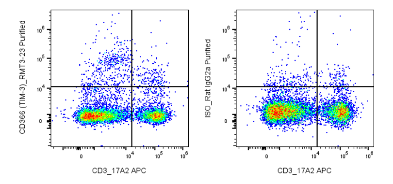
| Cat # | Size | Price | Quantity | |
|---|---|---|---|---|
| 205801 | 25 µg | $40 | ||
| 205802 | 100 µg | $95 |
| Clone | RMT3-23 |
|---|---|
| Application | Flow Cytometry, Block, IHC-F |
| Reactivity | Mouse |
| Format | Purified |
| Target Name | CD366, Tim-3, cell immunoglobulin and mucin domain containing 3 protein, hepatitis virus cellular receptor 2 |
| Isotype | Rat IgG2a |
| Antibody Type | Monoclonal |
| Regulatory Status | RUO |
| Formulation | Phosphate-buffered solution, pH 7.2, containing 0.09% sodium azide |
| Protein Concentration | 0.5 mg/mL |
| Storage & Handling | The antibody solution should be stored between 2°C and 8°C |
| Recommended Usage | For flow cytometric staining, it is recommended to use less than 0.2 µg of this reagent per 0.5-1.0 million cells in a 100 µL volume. Optimal reagent performance should be determined by titration for each specific application |
| See All Formats | Clone RMT3-23 |
Mouse CD336, also known as NKp46 and encoded by the Ncr1 gene, is a natural cytotoxicity receptor expressed almost exclusively on natural killer (NK) cells and a small subset of innate lymphoid cells (ILC1s and ILC3s). It is one of the principal activating receptors that enables NK cells to recognize and eliminate virus-infected, transformed, and stressed cells without prior antigen sensitization. Engagement of CD336 triggers NK cell activation, resulting in the release of cytotoxic granules containing perforin and granzymes, production of cytokines such as interferon-γ (IFN-γ) and tumor necrosis factor-α (TNF-α), and enhancement of immune responses through interactions with dendritic cells and macrophages.
Structurally, mouse CD336 is a type I transmembrane glycoprotein belonging to the immunoglobulin superfamily. It contains two extracellular C2-type immunoglobulin-like domains, a transmembrane region containing a positively charged residue, and a short cytoplasmic tail lacking intrinsic signaling motifs. Signal transduction occurs through association with the adaptor proteins FcRγ and CD3ζ, both of which contain immunoreceptor tyrosine-based activation motifs (ITAMs). CD336 recognizes a diverse range of ligands, including viral hemagglutinins from influenza and other viruses, fungal ligands, and several cellular molecules that are upregulated during cellular stress or malignant transformation. Additional tumor-associated ligands continue to be identified, although many remain incompletely characterized.
CD336 plays a critical role in host defense against viral infections, tumor immune surveillance, and regulation of inflammatory responses. Mice lacking CD336 exhibit impaired NK cell-mediated cytotoxicity and increased susceptibility to certain viral infections and experimental tumors. Conversely, excessive NK cell activation through CD336 may contribute to tissue injury during chronic inflammation. Because of its selective expression on NK cells, CD336 has become an important therapeutic target and biomarker. Anti-NKp46 antibodies are widely used to identify and isolate mouse NK cells, while preclinical therapeutic strategies include NKp46-directed bispecific antibodies, agonists that enhance NK cell activation, and engineered NK cell therapies designed to improve antitumor immunity. These approaches aim to potentiate innate immune responses while minimizing off-target effects on non-NK immune cells.
Rat IgG2a Isotype Control Antibody
Anti-Mouse CD366 (Tim-3) Antibody TDS
Have a product or application question? Consult our FAQs or contact us.