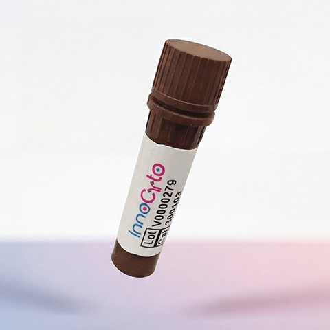
| Cat # | Size | Price | Quantity | |
|---|---|---|---|---|
| 207101 | 25 µg | $40 | ||
| 207102 | 100 µg | $95 |
| Clone | HMβ1-1 |
|---|---|
| Application | Flow Cytometry, IHC-F, IP, blocking |
| Reactivity | Mouse |
| Format | Purified |
| Target Name | CD29, Integrin β1 chain, VLA-β chain, gpIIa, ITGB1 |
| Isotype | Armenian Hamster IgG |
| Antibody Type | Monoclonal |
| Regulatory Status | RUO |
| Formulation | Phosphate-buffered solution, pH 7.2, containing 0.09% sodium azide |
| Protein Concentration | 0.5 mg/mL |
| Storage&Handling | The antibody solution should be stored between 2°C and 8°C |
| See All Formats | Clone HMβ1-1 |
CD29, also known as integrin β1 (ITGB1), is a ubiquitously expressed transmembrane adhesion receptor that forms the β1 subunit of at least 12 different integrin heterodimers through pairing with distinct α integrin subunits (including α1–α11 and αV). These heterodimers mediate cell adhesion to the extracellular matrix (ECM) and neighboring cells, regulating processes such as cell migration, proliferation, survival, differentiation, and tissue organization. CD29 is expressed on nearly all cell types, including leukocytes, epithelial cells, endothelial cells, fibroblasts, and stem cells, where it serves as a key mediator of tissue homeostasis and immune cell trafficking.
Structurally, CD29 is a type I transmembrane glycoprotein composed of a large extracellular domain, a single transmembrane helix, and a short cytoplasmic tail. The extracellular domain undergoes conformational changes that switch the receptor between inactive and high-affinity states, while the cytoplasmic tail interacts with adaptor proteins such as talin and kindlin to connect integrins to the actin cytoskeleton. CD29-containing integrins bind numerous extracellular ligands, including fibronectin, collagen, laminin, vitronectin, thrombospondin, and vascular cell adhesion molecule-1 (VCAM-1), depending on the associated α subunit. These interactions enable bidirectional signaling, whereby extracellular ligand binding influences intracellular signaling ("outside-in" signaling) and intracellular signals regulate ligand affinity ("inside-out" signaling).
Dysregulation of CD29 contributes to numerous diseases. Increased β1 integrin signaling promotes tumor growth, invasion, metastasis, resistance to chemotherapy, and survival of cancer stem cells. CD29 also plays important roles in fibrosis, chronic inflammation, autoimmune diseases, cardiovascular disease, and wound healing. In immune cells, β1 integrins regulate leukocyte adhesion and migration into inflamed tissues, making them important mediators of inflammatory responses.
Because of its central role in cell adhesion and signaling, CD29 has emerged as an important therapeutic target. Inhibitors of β1 integrins, blocking antibodies, peptides, and small molecules are being investigated to suppress tumor progression, reduce fibrosis, and limit inflammatory cell recruitment. Conversely, CD29 is widely used as a marker for mesenchymal stem cells and activated lymphocytes, and modulation of β1 integrin signaling is being explored to improve stem cell engraftment, tissue regeneration, and adoptive cell therapies such as CAR-T cell treatment.
Armenian Hamster IgG Isotype Control Antibody
Anti-Mouse/Rat CD29 Antibody TDS
Have a product or application question? Consult our FAQs or contact us.