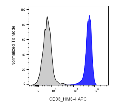
| Cat # | Size | Price | Quantity | |
|---|---|---|---|---|
| 114911 | 25 tests | $110 | ||
| 114912 | 100 tests | $250 |
| Clone | 033AM1 |
|---|---|
| Application | Flow Cytometry |
| Reactivity | Human |
| Format | APC |
| Target Name | CD33, Siglec-3,p67, gp67 |
| Isotype | Mouse IgG1 |
| Antibody Type | Monoclonal |
| Regulatory Status | RUO |
| Formulation | Phosphate-buffered solution, pH 7.2, containing 0.09% sodium azide and 0.2% (w/v) BSA |
| Protein Concentration | Supplied at a lot-specific concentration. |
| Storage & Handling | The antibody solution should be stored undiluted between 2°C and 8°C, and protected from prolonged exposure to light. Do not freeze. |
| Recommended Usage | For flow cytometric staining, it is recommended to use 5 µL of this reagent per 0.5-1.0 million cells in a 100 µL volume. Optimal reagent performance should be determined by titration for each specific application. APC has an excitation max at 650 nm and an emission max at 660 nm. |
| Excitation Laser | Red Laser (633 nm) |
| See All Formats | Clone 033AM1 |
CD33, also known as Siglec-3 (sialic acid-binding immunoglobulin-like lectin 3), is a transmembrane glycoprotein primarily expressed on cells of the myeloid lineage, including monocytes, macrophages, granulocytes, and myeloid progenitor cells. It functions as an inhibitory immune receptor that helps regulate innate immune responses and maintain immune homeostasis. CD33 contains an extracellular V-set immunoglobulin-like domain responsible for ligand recognition, a C2-set immunoglobulin-like domain, a single transmembrane region, and a cytoplasmic tail containing immunoreceptor tyrosine-based inhibitory motifs (ITIMs). Upon activation, these ITIMs recruit phosphatases such as SHP-1 and SHP-2, dampening cellular activation and inflammatory signaling.
The primary ligands for CD33 are sialylated glycans expressed on the surface of mammalian cells. By recognizing these self-associated carbohydrate structures, CD33 contributes to self-tolerance by suppressing unnecessary immune activation. Alterations in glycan expression can influence CD33 signaling and immune regulation.
CD33 is best known for its role in hematologic malignancies, particularly acute myeloid leukemia (AML), where it is expressed on leukemic blasts in the majority of patients while being absent from normal hematopoietic stem cells. This expression pattern has made CD33 an important therapeutic target. The antibody-drug conjugate gemtuzumab ozogamicin delivers a cytotoxic payload specifically to CD33-positive leukemic cells and has been approved for the treatment of certain patients with AML. Additional therapeutic strategies under investigation include bispecific antibodies, chimeric antigen receptor (CAR) T cells, CAR-natural killer (NK) cells, and other antibody-drug conjugates targeting CD33.
Beyond oncology, CD33 has been implicated in neurodegenerative disease. Genetic variants of CD33 are associated with altered risk of Alzheimer's disease, likely through effects on microglial function and amyloid-β clearance. Consequently, CD33 is also being explored as a therapeutic target for modulating neuroinflammation and enhancing microglial-mediated clearance of pathological proteins.
APC Mouse IgG1 Isotype Control Antibody
APC Anti-Human CD33 Antibody TDS
Have a product or application question? Consult our FAQs or contact us.