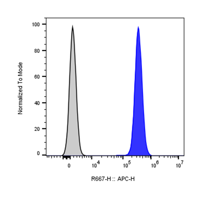
| Cat # | Size | Price | Quantity | |
|---|---|---|---|---|
| 112505 | 25 tests | $120 | ||
| 112506 | 100 tests | $250 |
| Clone | Her2AM1 |
|---|---|
| Application | Flow Cytometry |
| Reactivity | Human |
| Format | APC |
| Target Name | HER2, CD340, human epidermal growth factor receptor 2, Receptor tyrosine-protein kinase ErbB-2 (ERBB2) |
| Isotype | Mouse IgG1 |
| Antibody Type | Monoclonal |
| Regulatory Status | RUO |
| Formulation | Phosphate-buffered solution, pH 7.2, containing 0.09% sodium azide and 0.2% (w/v) BSA |
| Protein Concentration | Supplied at a lot-specific concentration. |
| Storage&Handling | The antibody solution should be stored undiluted between 2°C and 8°C, and protected from prolonged exposure to light. Do not freeze. |
| Recommended Usage | For flow cytometric staining, it is recommended to use 5 µL of this reagent per 0.5-1.0 million cells in a 100 µL volume. Optimal reagent performance should be determined by titration for each specific application. APC has an excitation max at 650 nm and an emission max at 660 nm. |
| Excitation Laser | Red Laser (633 nm) |
| Research Areas | Mesenchymal Stem Cells, Embryogenesis, Cancer Marker, Cancer Immunology |
| See All Formats | Clone Her2AM1 |
CD340, more commonly known as HER2 (human epidermal growth factor receptor 2) or ErbB2, is a member of the ErbB family of receptor tyrosine kinases. It plays a critical role in regulating cell growth, survival, differentiation, and repair. Unlike other members of its family, CD340 does not have a known direct ligand; instead, it functions primarily through heterodimerization with other ErbB receptors such as EGFR (ErbB1), HER3 (ErbB3), or HER4 (ErbB4). This dimerization activates intracellular signaling pathways, including the PI3K/AKT and MAPK pathways, which promote cell proliferation and inhibit apoptosis.
Structurally, CD340 is a transmembrane glycoprotein composed of three main domains: an extracellular ligand-binding domain, a single hydrophobic transmembrane region, and an intracellular tyrosine kinase domain. The extracellular region contains four subdomains responsible for receptor dimerization, while the intracellular domain mediates downstream signaling through autophosphorylation of tyrosine residues.
Although CD340 lacks a direct ligand, its activity is tightly regulated through interactions with other ErbB receptors and their ligands, such as epidermal growth factor (EGF) and neuregulins. Overexpression or amplification of the CD340 gene leads to constitutive activation of signaling pathways, which is strongly associated with oncogenesis.
CD340 is most notably implicated in breast cancer, where HER2 overexpression occurs in approximately 15–20% of cases and is associated with aggressive tumor growth and poor prognosis. It is also involved in other cancers, including gastric and ovarian cancers.
Therapeutically, CD340 is a major target in oncology. Monoclonal antibodies such as trastuzumab and pertuzumab, as well as small molecule inhibitors like lapatinib, have been developed to inhibit HER2 signaling. These targeted therapies have significantly improved outcomes in HER2-positive cancers, making CD340 a cornerstone of precision medicine.
APC Mouse IgG1 Isotype Control Antibody
APC Anti-human CD340 (ErbB2/HER-2) Antibody TDS
Have a product or application question? Consult our FAQs or contact us.