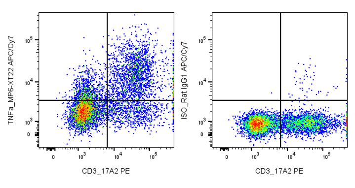
| Cat # | Size | Price | Quantity | |
|---|---|---|---|---|
| 203303 | 25 tests | $80 | ||
| 203304 | 100 tests | $150 |
| Clone | MP6-XT22 |
|---|---|
| Application | Intracellular Flow Cytometry |
| Reactivity | Mouse |
| Format | APC/Cyanine7 |
| Target Name | TNF-α, TNF-alpha, Tumor necrosis factor-α, Macrophage cytotoxic factor (MCF) |
| Isotype | Rat IgG1 |
| Antibody Type | Monoclonal |
| Regulatory Status | RUO |
| Formulation | Phosphate-buffered solution, pH 7.2, containing 0.09% sodium azide and 0.2% (w/v) BSA |
| Protein Concentration | Supplied at a lot-specific concentration. |
| Storage&Handling | The antibody solution should be stored undiluted between 2°C and 8°C, and protected from prolonged exposure to light. Do not freeze. |
| Recommended Usage | For flow cytometric staining, it is recommended to use 5 uL of this reagent per 0.5-1.0 million cells in a 100 µL volume. Optimal reagent performance should be determined by titration for each specific application. APC/Cyanine7 has an excitation max at 650 nm and an emission max at 774 nm. |
| Excitation Laser | Red Laser (633 nm) |
| Isotype Controls | 303412 |
| Antibody Family | Mouse Antibodies |
| Research Areas | Cytokines, Monocytes, Macrophages, Neutrophils, T cells, Inflammation, Autoimmunity |
| See All Formats | Clone MP6-XT22 |
Mouse tumor necrosis factor-alpha (TNF-α) is a pro-inflammatory cytokine produced primarily by activated macrophages, T cells, and other immune cells. It plays a central role in innate and adaptive immunity by regulating inflammation, cell survival, apoptosis, and immune cell activation. TNF-α is rapidly induced in response to infection or tissue injury and helps coordinate host defense by promoting cytokine production, leukocyte recruitment, and pathogen clearance.
Structurally, TNF-α is initially synthesized as a type II transmembrane protein that can be cleaved by metalloproteases such as TACE (ADAM17) to release a soluble form. Both membrane-bound and soluble TNF-α function as homotrimers, which are the biologically active forms. TNF-α exerts its effects by binding to two receptors, TNFR1 (p55) and TNFR2 (p75), which differ in expression patterns and downstream signaling pathways.
The primary ligands for TNF receptors are TNF-α itself and the related cytokine lymphotoxin-α. Engagement of TNFR1 often leads to activation of NF-κB and MAPK pathways or induction of apoptosis, while TNFR2 is more associated with immune regulation and cell survival.
In disease, dysregulated TNF-α production contributes to chronic inflammatory and autoimmune conditions in mouse models, including arthritis, colitis, and sepsis. Therapeutically, TNF-α is a major target in inflammatory disease, and anti-TNF biologics have been widely developed. In mice, TNF modulation is also used experimentally to study inflammation, cancer immunity, and immune-mediated pathology.
APC/Cyanine7 Anti-Mouse TNF-α Antibody TDS
Intracellular Flow Cytometry Protocol
Have a product or application question? Consult our FAQs or contact us.