| Cat # | Size | Price | Quantity | |
|---|---|---|---|---|
| 809803 | 25 ug | $245 | ||
| 809804 | 100 ug | $595 |
| Application | ELISA, BLI |
|---|---|
| Format | Liquid, Biotinylated |
| Expression Host | CHO |
| Target Name | CD73, NT5E |
| Species | Human |
| Sources | Recombinant Human CD73 Protein (Trp27-Lys547) with C-terminus His-Avi-tag is expressed in CHO cell. This protein was site-specifically labeled with Biotin by BirA ligase. |
| Accession Number | P21589 |
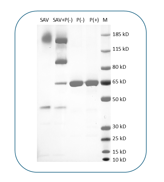
| Molecular Weight | The protein has a predicted molecular weight of 61.3 kDa. Under DTT-reducing conditions, it migrates at approximately 65 kDa on SDS-PAGE. |
| Affinity Tag | C-His-Avi |
| Purity | >95% based on SDS-PAGE under reducing condition |
| Regulatory Status | RUO |
| Formulation | 1xPBS buffer, pH7.4, 0.22 µm filtered |
| Endotoxin level | Not tested |
| Protein Concentration | 25µg size is bottled at 0.2mg/mL concentration. 100 µg size is supplied at a lot-specific concentration. |
| Storage and Handling | Briefly centrifuge the vial upon receipt. An unopened vial can be stored at 4°C for up to 2 weeks, or at -20°C or below for up to six months. The protein may be further diluted to 0.1 mg/mL using 0.22 µm-filtered PBS buffer (pH 7.4). For long-term storage, the diluted stock solution should be aliquoted and stored at ≤ –70°C to minimize freeze-thaw cycles. If additional dilution is required, carrier proteins such as FBS or BSA should be added to maintain protein stability. |
| Recommended Usage | For detection, use a secondary reagent with this product. |
| Research Areas | Endothelial cells, T cells, B cells, Tregs, Macrophages, Cancer Marker, Immunosuppression, Angiogenesis |
CD73, also known as ecto-5'-nucleotidase (NT5E), is a glycosylphosphatidylinositol (GPI)-anchored cell surface enzyme that plays a critical role in purinergic signaling and immune regulation. CD73 catalyzes the extracellular conversion of adenosine monophosphate (AMP) to adenosine and inorganic phosphate, representing the final step in the generation of extracellular adenosine. This enzymatic activity is crucial for regulating immune responses, as adenosine acts as a potent immunosuppressive molecule that dampens T cell activation, reduces inflammatory cytokine production, and promotes regulatory T cell function. CD73 is expressed on various cell types, including lymphocytes, endothelial cells, epithelial cells, and many tumor cells, where it contributes to maintaining tissue homeostasis and modulating immune responses.
Structurally, CD73 is a homodimeric metalloenzyme of approximately 70 kDa per monomer, anchored to the cell membrane via a GPI linkage. Each monomer contains two α/β domains that form the catalytic core, with the active site located at the interface between these domains. The enzyme requires metal ions, particularly zinc, for catalytic activity. The catalytic mechanism involves the coordination of AMP substrate within a pocket formed by conserved residues, followed by hydrolysis of the phosphate ester bond. The GPI anchor localizes CD73 to lipid rafts on the cell surface, positioning it optimally for interaction with other signaling molecules and facilitating its role in cell-cell communication.
The primary substrate for CD73 is AMP, which is converted to adenosine. The adenosine produced then acts as a ligand for adenosine receptors (A1, A2A, A2B, and A3) on nearby cells, mediating diverse physiological effects. CD73 works in concert with CD39 (ectonucleoside triphosphate diphosphohydrolase-1), which converts ATP and ADP to AMP, forming the CD39-CD73 axis that is central to adenosine generation in the extracellular space.
In disease contexts, CD73 plays a significant role in cancer progression and immune evasion. Many tumors overexpress CD73, creating an adenosine-rich immunosuppressive microenvironment that inhibits antitumor immunity and promotes tumor growth, angiogenesis, and metastasis. High CD73 expression correlates with poor prognosis in various cancers, including triple-negative breast cancer, melanoma, ovarian cancer, and colorectal cancer. Therapeutically, CD73 has emerged as an important immune checkpoint target. Multiple CD73 inhibitors and blocking antibodies are in clinical development, including oleclumab and quemliclustat, which aim to reduce adenosine production and restore antitumor immune responses. These agents are being tested both as monotherapies and in combination with other immune checkpoint inhibitors such as anti-PD-1/PD-L1 antibodies, showing promise in enhancing cancer immunotherapy efficacy. Additionally, CD73 inhibition is being explored for treating fibrotic diseases and ischemia-reperfusion injury, establishing it as a versatile therapeutic target across multiple disease contexts.
Biotin Human CD73 Protein (C-His-Avi) TDS
Human CD73 Protein (C-His-Avi)
APC Human CD73 Protein (C-His)
Have a product or application question? Consult our FAQs or contact us.