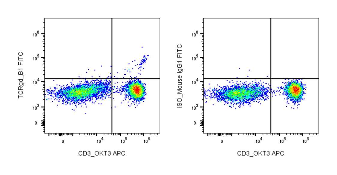
| Cat # | Size | Price | Quantity | |
|---|---|---|---|---|
| 111903 | 25 tests | $75 | ||
| 111904 | 100 tests | $170 |
| Clone | B1 |
|---|---|
| Application | Flow Cytometry |
| Reactivity | Human |
| Format | FITC |
| Target Name | T cell receptor γ/δ, γ/δ TCR, TCR-γ/δ |
| Isotype | Mouse IgG1 |
| Antibody Type | Monoclonal |
| Regulatory Status | RUO |
| Formulation | Phosphate-buffered solution, pH 7.2, containing 0.09% sodium azide and 0.2% (w/v) BSA |
| Protein Concentration | Supplied at a lot-specific concentration. |
| Storage&Handling | The antibody solution should be stored undiluted between 2°C and 8°C, and protected from prolonged exposure to light. Do not freeze. |
| Recommended Usage | For flow cytometric staining, it is recommended to use 5 µL of this reagent per 0.5-1.0 million cells in a 100 µL volume. Optimal reagent performance should be determined by titration for each specific application. FITC has an excitation max at 493 nm and an emission max at 525 nm. |
| Excitation Laser | Blue Laser (488 nm) |
| Research Areas | T cells, T cell Activation, Adaptive Immunity |
| See All Formats | Clone B1 |
Human TCR γ/δ refers to the T cell receptor expressed on γδ T cells, a distinct subset of T lymphocytes that bridge innate and adaptive immunity. Unlike conventional αβ T cells, γδ T cells respond rapidly to stress signals, infection, and cellular transformation without requiring classical antigen presentation. They are enriched in epithelial and mucosal tissues, where they contribute to immune surveillance, tissue repair, and early defense against pathogens.
Structurally, the γδ T cell receptor is a heterodimer composed of one gamma (γ) chain and one delta (δ) chain, each containing variable (V), diversity (D, for δ), joining (J), and constant (C) regions generated through somatic recombination. The receptor associates with the CD3 signaling complex, which transduces activation signals into the cell. Compared to αβ TCRs, γδ TCRs exhibit more limited diversity but greater flexibility in antigen recognition.
The ligands recognized by γδ TCRs are diverse and not restricted to peptide antigens presented by classical major histocompatibility complex (MHC) molecules. Instead, they detect non-peptide phosphoantigens, lipid antigens, and stress-induced molecules such as MICA/MICB and butyrophilin family members (e.g., BTN3A1). This allows γδ T cells to sense metabolic dysregulation and cellular stress commonly associated with infection or malignancy.
In disease, γδ T cells play complex roles. They contribute to protective immunity against infections and tumors by producing cytokines and exerting cytotoxic activity. However, they can also promote inflammation in autoimmune diseases such as psoriasis and multiple sclerosis. Dysregulation of γδ T cell responses may therefore contribute to both protective and pathological outcomes.
Therapeutically, γδ T cells are being actively explored in immunotherapy. Their ability to recognize tumor cells in an MHC-independent manner makes them attractive candidates for cancer treatment. Approaches include adoptive transfer of expanded γδ T cells, stimulation with phosphoantigens, and engineering γδ TCR-based therapies. These strategies aim to harness their rapid response and broad reactivity to enhance anti-tumor immunity while minimizing the risk of immune escape.
FITC Mouse IgG1 Isotype Control Antibody
FITC Anti-Human TCR γ/δ Antibody TDS
Have a product or application question? Consult our FAQs or contact us.