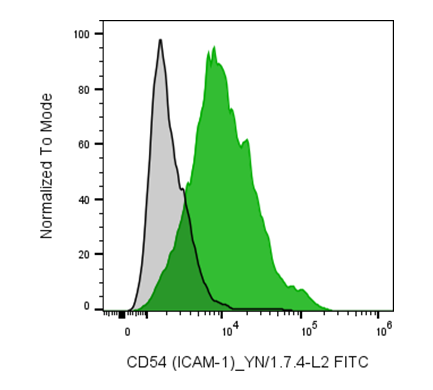
| Cat # | Size | Price | Quantity | |
|---|---|---|---|---|
| 206807 | 25 tests | $40 | ||
| 206808 | 100 tests | $80 |
| Clone | YN1/1.7.4 |
|---|---|
| Application | Flow Cytometry |
| Reactivity | Mouse |
| Format | FITC |
| Target Name | CD54, ICAM-1, Ly-47 |
| Isotype | Rat IgG2b |
| Antibody Type | Monoclonal |
| Regulatory Status | RUO |
| Formulation | Phosphate-buffered solution, pH 7.2, containing 0.09% sodium azide and 0.2% (w/v) BSA |
| Protein Concentration | Supplied at a lot-specific concentration. |
| Storage&Handling | The antibody solution should be stored undiluted between 2°C and 8°C, and protected from prolonged exposure to light. Do not freeze. |
| Recommended Usage | For flow cytometric staining, it is recommended to use 5 uL of this reagent per 0.5-1.0 million cells in a 100 µL volume. Optimal reagent performance should be determined by titration for each specific application. FITC has an excitation max at 493 nm and an emission max at 525 nm. |
| Excitation Laser | Blue Laser (488 nm) Green/Yellow laser (532/561nm) |
| See All Formats | Clone YN1/1.7.4 |
Mouse CD54, also known as intercellular adhesion molecule-1 (ICAM-1), is a transmembrane glycoprotein encoded by the Icam1 gene and is a member of the immunoglobulin superfamily. It is constitutively expressed at low levels on endothelial cells, epithelial cells, fibroblasts, and many leukocytes, but is strongly upregulated by inflammatory cytokines such as tumor necrosis factor-α (TNF-α), interleukin-1β (IL-1β), and interferon-γ (IFN-γ). CD54 plays a central role in immune surveillance by mediating leukocyte adhesion to vascular endothelium and promoting migration of immune cells into inflamed tissues.
Structurally, mouse CD54 consists of five extracellular immunoglobulin-like domains, a single transmembrane domain, and a short cytoplasmic tail. Although the cytoplasmic domain lacks intrinsic enzymatic activity, it participates in intracellular signaling through interactions with cytoskeletal and adaptor proteins. The principal ligands for CD54 are the β2 integrins lymphocyte function-associated antigen-1 (LFA-1; CD11a/CD18) and macrophage-1 antigen (Mac-1; CD11b/CD18), which are expressed on leukocytes. These interactions stabilize immune cell adhesion, facilitate antigen presentation, and support T cell activation.
CD54 has been implicated in numerous inflammatory and autoimmune diseases in mouse models, including experimental autoimmune encephalomyelitis, rheumatoid arthritis, inflammatory bowel disease, and transplant rejection. Elevated CD54 expression also contributes to leukocyte infiltration during infection and tissue injury. Conversely, reduced CD54 function can impair effective immune responses. In cancer, CD54 expression may either enhance antitumor immunity by promoting immune cell interactions or facilitate tumor progression depending on the tumor microenvironment.
Because of its central role in leukocyte trafficking and immune activation, CD54 is an attractive therapeutic target. Blocking CD54 or its interaction with LFA-1 can reduce excessive inflammation in preclinical models, while strategies that increase CD54 expression on tumors may improve immune-mediated tumor killing. Consequently, mouse CD54 is widely used in studies investigating inflammatory disease mechanisms and the development of immunomodulatory therapies.
FITC Rat IgG2b Isotype Control
FITC Anti-Mouse CD54 (ICAM-1) Antibody TDS
Have a product or application question? Consult our FAQs or contact us.