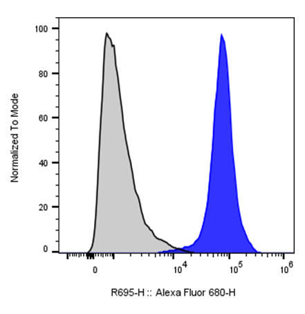
| Cat # | Size | Price | Quantity | |
|---|---|---|---|---|
| 113603 | 25 tests | $135 | ||
| 113604 | 100 tests | $280 |
| Clone | 112AMg1 |
|---|---|
| Application | Flow Cytometry |
| Reactivity | Human |
| Format | iF647 |
| Target Name | CD112, Nectin-2, Poliovirus Receptor Related 2 Protein (PRR2), Hve B |
| Isotype | Mouse IgG1 |
| Antibody Type | Monoclonal |
| Regulatory Status | RUO |
| Formulation | Phosphate-buffered solution, pH 7.2, containing 0.09% sodium azide and 0.2% (w/v) BSA |
| Protein Concentration | Supplied at a lot-specific concentration. |
| Storage&Handling | The antibody solution should be stored undiluted between 2°C and 8°C, and protected from prolonged exposure to light. Do not freeze. |
| Recommended Usage | For flow cytometric staining, it is recommended to use 5 µL of this reagent per 0.5-1.0 million cells in a 100 µL volume. Optimal reagent performance should be determined by titration for each specific application. iF647 has an excitation max at 656 nm and an emission max at 670 nm. |
| Excitation Laser | Red Laser (633 nm) |
| Isotype Controls | 301413 |
| Antibody Family | Human Antibodies |
| Research Areas | Adhesion Molecules, Angiogenesis, Epithelial cells, Endothelial cells, Dendritic cells, Cancer Marker, Immune Checkpoints |
| See All Formats | Clone 112AMg1 |
Human CD112, also known as nectin-2 or poliovirus receptor-related protein 2 (PVRL2), is a cell adhesion molecule belonging to the immunoglobulin superfamily. It is broadly expressed on epithelial cells, endothelial cells, and antigen-presenting cells. CD112 plays a dual role in maintaining cell–cell adhesion and regulating immune responses, particularly in interactions between immune cells and target tissues.
Structurally, CD112 is a type I transmembrane glycoprotein composed of three extracellular immunoglobulin-like domains (one V-type followed by two C2-type domains), a single transmembrane region, and a short cytoplasmic tail. This structure enables it to participate in both homophilic (CD112–CD112) and heterophilic interactions with other nectin family members.
Functionally, CD112 serves as a ligand for several immune receptors, most notably CD226 (DNAM-1), TIGIT, and CD112R (PVRIG). Binding to CD226 provides co-stimulatory signals that enhance T cell and natural killer (NK) cell activation, while interactions with TIGIT and CD112R deliver inhibitory signals that dampen immune responses. Thus, CD112 is part of a regulatory axis balancing immune activation and inhibition.
In disease, CD112 is frequently upregulated in various cancers, where it contributes to immune evasion by preferentially engaging inhibitory receptors such as TIGIT and CD112R on T cells and NK cells. This suppresses anti-tumor immunity and supports tumor progression.
Therapeutically, CD112 is an emerging target in cancer immunotherapy. Strategies include blocking inhibitory pathways (e.g., TIGIT or CD112R antibodies) or enhancing CD226-mediated activation. These approaches aim to restore immune function and improve anti-tumor responses, often in combination with other checkpoint inhibitors.
iF647 Anti-human CD112 (Nectin-2) Antibody TDS
Have a product or application question? Consult our FAQs or contact us.