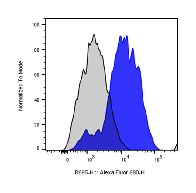
| Cat # | Size | Price | Quantity | |
|---|---|---|---|---|
| 113403 | 25 tests | $135 | ||
| 113404 | 100 tests | $280 |
| Clone | 3A6 |
|---|---|
| Application | Flow Cytometry |
| Reactivity | Human |
| Format | iF647 |
| Target Name | CD166, CD6 ligand, Activated Leukocyte Cell Adhesion Molecule, ALCAM |
| Isotype | Mouse IgG1 |
| Antibody Type | Monoclonal |
| Regulatory Status | RUO |
| Formulation | Phosphate-buffered solution, pH 7.2, containing 0.09% sodium azide and 0.2% (w/v) BSA |
| Protein Concentration | Supplied at a lot-specific concentration. |
| Storage&Handling | The antibody solution should be stored undiluted between 2°C and 8°C, and protected from prolonged exposure to light. Do not freeze. |
| Recommended Usage | For flow cytometric staining, it is recommended to use 5 µL of this reagent per 0.5-1.0 million cells in a 100 µL volume. Optimal reagent performance should be determined by titration for each specific application. iF647 has an excitation max at 656 nm and an emission max at 670 nm. |
| Excitation Laser | Red Laser (633 nm) |
| Research Areas | Adhesion Molecules, Mesenchymal Stem cells, Activated T cells, B cells, Monocytes, Dendritic Cells, Vascular Endothelial cells, Cancer Marker, Angiogenesis |
| See All Formats | Clone 3A6 |
CD166, also known as activated leukocyte cell adhesion molecule (ALCAM), is a cell surface glycoprotein involved in cell–cell adhesion and immune regulation. It is broadly expressed on a variety of cell types, including activated T cells, monocytes, endothelial cells, epithelial cells, and certain stem and progenitor cells. CD166 plays an important role in mediating interactions between immune cells and between immune and non-immune cells, contributing to processes such as leukocyte trafficking, immune synapse formation, and tissue organization.
Structurally, CD166 is a type I transmembrane protein belonging to the immunoglobulin (Ig) superfamily. It contains five extracellular Ig-like domains (two variable-like and three constant-like domains), a single transmembrane region, and a short cytoplasmic tail. The extracellular domains mediate both homophilic interactions (CD166–CD166) and heterophilic binding, enabling flexible adhesion functions in different cellular contexts.
The principal ligand for CD166 is CD6, a receptor expressed on T cells. The CD166–CD6 interaction is critical for stabilizing the immunological synapse and enhancing T cell activation and proliferation. In addition to this heterophilic binding, CD166 can also engage in homophilic interactions, which contribute to cell aggregation and tissue integrity.
In disease, CD166 is implicated in cancer progression, inflammation, and autoimmune disorders. It is frequently overexpressed in a variety of malignancies, including colorectal, breast, prostate, and melanoma, where its expression is often associated with tumor invasion, metastasis, and poor prognosis. CD166 is also considered a marker of cancer stem cells in certain tumors. In inflammatory diseases, dysregulated CD166 expression may influence immune cell activation and tissue infiltration.
Therapeutically, CD166 is being investigated as a target for cancer treatment and immune modulation. Strategies include monoclonal antibodies, antibody-drug conjugates, and CAR T cell approaches aimed at eliminating CD166-expressing tumor cells. Additionally, modulating CD166–CD6 interactions may provide a means to regulate T cell responses in autoimmune diseases and transplantation. Ongoing research continues to explore its potential as both a biomarker and therapeutic target.
iF647 Mouse IgG1 Isotype Control Antibody
iF647 Anti-Human CD166 Antibody TDS
Have a product or application question? Consult our FAQs or contact us.