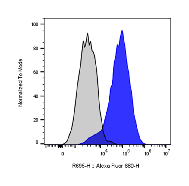
| Cat # | Size | Price | Quantity | |
|---|---|---|---|---|
| 112703 | 25 tests | $120 | ||
| 112704 | 100 tests | $270 |
| Clone | HLAGAR1 |
|---|---|
| Application | Flow Cytometry |
| Reactivity | Human |
| Format | iF647 |
| Target Name | HLA-G, Human Leukocyte Antigen-G |
| Isotype | Rat IgG1 |
| Antibody Type | Monoclonal |
| Regulatory Status | RUO |
| Formulation | Phosphate-buffered solution, pH 7.2, containing 0.09% sodium azide and 0.2% (w/v) BSA |
| Protein Concentration | Supplied at a lot-specific concentration. |
| Storage&Handling | The antibody solution should be stored undiluted between 2°C and 8°C, and protected from prolonged exposure to light. Do not freeze. |
| Recommended Usage | For flow cytometric staining, it is recommended to use 5 µL of this reagent per 0.5-1.0 million cells in a 100 µL volume. Optimal reagent performance should be determined by titration for each specific application. iF647 has an excitation max at 656 nm and an emission max at 670 nm. |
| Excitation Laser | Red Laser (633 nm) |
| Research Areas | Immune Checkpoints, Cancer Marker, Infectious Diseases, Autoimmunity |
| See All Formats | Clone HLAGAR1 |
HLA-G is a non-classical major histocompatibility complex (MHC) class I molecule encoded within the human leukocyte antigen (HLA) region. Unlike classical MHC class I proteins, HLA-G has limited polymorphism and a restricted tissue distribution, primarily expressed at the maternal–fetal interface, where it plays a critical role in immune tolerance during pregnancy.
Structurally, HLA-G consists of a heavy chain associated with β2-microglobulin and presents peptides similarly to other MHC class I molecules. However, alternative splicing generates multiple isoforms, including both membrane-bound (e.g., HLA-G1) and soluble forms (e.g., HLA-G5). These structural variants contribute to its diverse immunomodulatory functions.
HLA-G interacts with inhibitory receptors such as ILT2 (LILRB1), ILT4 (LILRB2), and KIR2DL4 expressed on immune cells including natural killer (NK) cells, T cells, and antigen-presenting cells. Through these ligand–receptor interactions, HLA-G suppresses immune responses by inhibiting cytotoxic activity, reducing cytokine production, and promoting regulatory cell phenotypes.
In disease contexts, aberrant expression of HLA-G has been associated with cancer, viral infections, and autoimmune disorders. Many tumors exploit HLA-G expression to evade immune surveillance, leading to poorer clinical outcomes. Conversely, reduced HLA-G expression may contribute to pregnancy complications such as preeclampsia or recurrent miscarriage.
Therapeutically, HLA-G represents a promising target in both immunosuppression and immuno-oncology. Enhancing HLA-G activity could be beneficial in transplantation and autoimmune diseases by promoting immune tolerance. In contrast, blocking HLA-G or its receptors is being explored as a strategy to restore anti-tumor immunity. Ongoing research aims to better understand its mechanisms and develop targeted therapies that modulate HLA-G pathways.
iF647 Rat IgG1 Isotype Control Antibody
iF647 Anti-human HLA-G Antibody TDS
Have a product or application question? Consult our FAQs or contact us.