| Cat # | Size | Price | Quantity | |
|---|---|---|---|---|
| 203903 | 25 tests | $100 | ||
| 203904 | 100 tests | $230 |
| Clone | 1B1 |
|---|---|
| Application | Flow Cytometry |
| Reactivity | Mouse |
| Format | iF647 |
| Target Name | CD1d, CD1, CD1.1, Ly-38 |
| Isotype | Rat IgG2b |
| Antibody Type | Monoclonal |
| Regulatory Status | RUO |
| Formulation | Phosphate-buffered solution, pH 7.2, containing 0.09% sodium azide and 0.2% (w/v) BSA |
| Protein Concentration | Supplied at a lot-specific concentration. |
| Storage&Handling | The antibody solution should be stored undiluted between 2°C and 8°C, and protected from prolonged exposure to light. Do not freeze. |
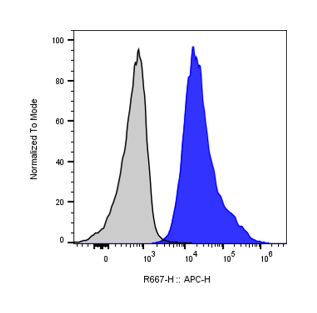
| Recommended Usage | For flow cytometric staining, it is recommended to use 5 µL of this reagent per 0.5-1.0 million cells in a 100 µL volume. Optimal reagent performance should be determined by titration for each specific application. iF647 has an excitation max at 656 nm and an emission max at 670 nm. |
| Excitation Laser | Red Laser (633 nm) |
| Research Areas | Antigen Presentation, iNKT, Autoimmunity, Cancer Immunology |
| See All Formats | Clone 1B1 |
CD1d is a non-classical antigen-presenting molecule that belongs to the CD1 family of glycoproteins, which are structurally related to major histocompatibility complex (MHC) class I molecules. Unlike classical MHC molecules that present peptide antigens, CD1d specializes in presenting lipid and glycolipid antigens to a unique subset of T cells known as invariant natural killer T (iNKT) cells. CD1d is expressed on a variety of antigen-presenting cells, including dendritic cells, macrophages, B cells, and certain epithelial cells. Through its interaction with NKT cells, CD1d plays an important role in bridging innate and adaptive immunity.
Structurally, CD1d consists of a heavy chain associated with β2-microglobulin, similar to MHC class I molecules. The heavy chain forms a deep hydrophobic binding groove composed of two pockets (commonly referred to as the A′ and F′ pockets) that accommodate the lipid tails of antigens. The hydrophilic head groups of these lipid molecules protrude from the binding groove and are recognized by the T cell receptor (TCR) on NKT cells. This structural configuration enables CD1d to present a wide variety of lipid-based antigens derived from both self and microbial sources.
CD1d binds several classes of lipid ligands. Endogenous ligands include self-glycosphingolipids and phospholipids that help regulate NKT cell homeostasis. Exogenous ligands often originate from microbes, including glycolipids from bacteria such as Sphingomonas species. One of the best-known synthetic ligands is α-galactosylceramide (α-GalCer), originally derived from a marine sponge-associated bacterium. When presented by CD1d, α-GalCer strongly activates iNKT cells, leading to rapid production of cytokines such as interferon-γ and interleukin-4.
Through activation of NKT cells, CD1d plays a role in numerous immune-mediated conditions. It has been implicated in cancer immunity, infectious diseases, autoimmune disorders, and inflammatory diseases. In cancer, CD1d-mediated activation of NKT cells can enhance anti-tumor immune responses by stimulating cytotoxic lymphocytes and promoting cytokine release. Conversely, dysregulated CD1d–NKT interactions may contribute to autoimmune diseases such as type 1 diabetes, inflammatory bowel disease, and systemic lupus erythematosus.
Because of its central role in regulating NKT cell responses, CD1d has become an attractive therapeutic target. Strategies under investigation include lipid-based agonists that activate NKT cells to boost anti-tumor immunity, modified glycolipid ligands designed to bias cytokine responses, and antibody-based approaches that modulate CD1d function. In addition, CD1d-restricted antigen presentation is being explored in vaccine design and immunotherapy platforms aimed at harnessing the rapid immunoregulatory properties of NKT cells. These approaches highlight CD1d as an important molecule at the intersection of lipid antigen presentation and immune modulation.
iF647 Rat IgG2b Isotype Control Antibody
iF647 Anti-mouse CD1d (CD1.1, Ly-38) Antibody TDS
PE Anti-Mouse H-2 Antibody, Clone M1/42
iF647 Anti-mouse I-A/I-E Antibody, Clone M5/114.15.2
Have a product or application question? Consult our FAQs or contact us.