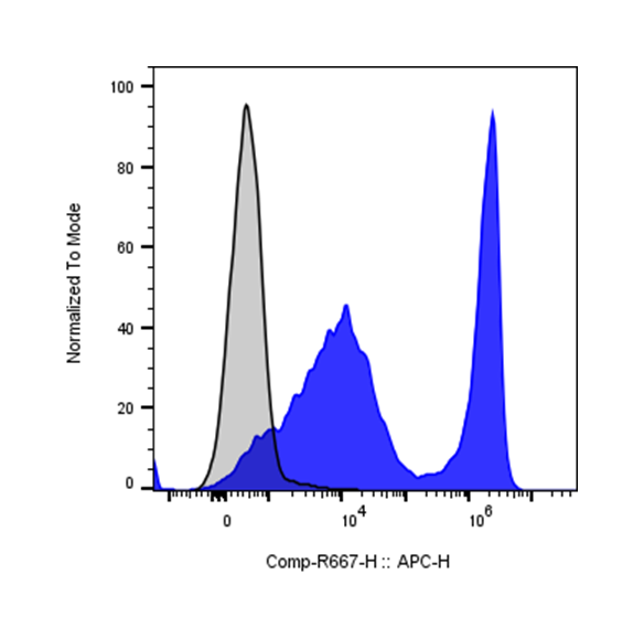
| Cat # | Size | Price | Quantity | |
|---|---|---|---|---|
| 203603 | 25 tests | $80 | ||
| 203604 | 100 tests | $180 |
| Clone | 30-H12 |
|---|---|
| Application | Flow Cytometry |
| Reactivity | Mouse |
| Format | iF647 |
| Target Name | CD90.2, Thy1.2, Thy-1.2 |
| Isotype | Rat IgG2b |
| Antibody Type | Monoclonal |
| Regulatory Status | RUO |
| Formulation | Phosphate-buffered solution, pH 7.2, containing 0.09% sodium azide and 0.2% (w/v) BSA |
| Protein Concentration | Supplied at a lot-specific concentration. |
| Storage&Handling | The antibody solution should be stored undiluted between 2°C and 8°C, and protected from prolonged exposure to light. Do not freeze. |
| Recommended Usage | For flow cytometric staining, it is recommended to use 5 µL of this reagent per 0.5-1.0 million cells in a 100 µL volume. Optimal reagent performance should be determined by titration for each specific application. iF647 has an excitation max at 656 nm and an emission max at 670 nm. |
| Excitation Laser | Red Laser (633 nm) |
| Research Areas | T cells, Hematopoietic Stem Cells, Neurons |
| See All Formats | Clone 30-H12 |
Mouse CD90.2, also known as Thy-1.2, is a glycosylphosphatidylinositol (GPI)-anchored cell surface glycoprotein that belongs to the immunoglobulin superfamily. It is widely expressed in mice on several cell types, including thymocytes, mature T lymphocytes, neurons, fibroblasts, and some stem or progenitor cells. In immunology research, CD90.2 serves as an important marker for identifying mouse T cells and distinguishing mouse strains that express the Thy-1.2 allele from those expressing the alternative Thy-1.1 allele.
Structurally, CD90.2 is a relatively small protein of approximately 25–37 kDa depending on glycosylation. It contains a single extracellular immunoglobulin-like domain, multiple glycosylation sites, and is attached to the outer leaflet of the plasma membrane through a GPI anchor rather than a transmembrane region. Because it lacks an intracellular domain, CD90.2 does not signal directly but instead participates in signaling through associations with other membrane proteins and lipid raft microdomains. This arrangement allows CD90.2 to influence cell–cell interactions and intracellular signaling pathways indirectly.
Although the precise physiological ligands of CD90 are not completely defined, studies have shown that Thy-1 molecules can interact with integrins, such as αvβ3 integrin, on neighboring cells. These interactions contribute to cell adhesion, communication between immune cells and stromal cells, and regulation of cell activation and migration.
CD90 plays roles in immune regulation, neuronal development, and tissue repair. Abnormal CD90 expression has been associated with inflammatory conditions, fibrosis, and certain cancers, where it may influence cell proliferation and tumor–stromal interactions. In therapeutic research, CD90 is used as a marker to isolate specific cell populations, including T cells and stem cells. Antibodies against CD90.2 are widely used in mouse models for immune cell depletion, cell tracking, and transplantation studies.
iF647 Rat IgG2b Isotype Control
iF647 Anti-Mouse CD90.2 (Thy1.2) Antibody TDS
Have a product or application question? Consult our FAQs or contact us.