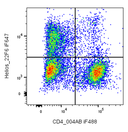
| Cat # | Size | Price | Quantity | |
|---|---|---|---|---|
| 204603 | 25 tests | $165 | ||
| 204604 | 100 tests | $320 |
| Clone | 22F6 |
|---|---|
| Application | Intracellular Flow Cytometry |
| Reactivity | Mouse, Human |
| Format | iF647 |
| Target Name | Helios, IKAROS family zinc finger 2, IKZF2, ZNF1A2, ZNFN1A2 |
| Isotype | Armenian Hamster IgG |
| Antibody Type | Monoclonal |
| Regulatory Status | RUO |
| Formulation | Phosphate-buffered solution, pH 7.2, containing 0.09% sodium azide and 0.2% (w/v) BSA |
| Protein Concentration | Supplied at a lot-specific concentration. |
| Storage&Handling | The antibody solution should be stored undiluted between 2°C and 8°C, and protected from prolonged exposure to light. Do not freeze. |
| Recommended Usage | For flow cytometric staining, it is recommended to use 5 uL of this reagent per 0.5-1.0 million cells in a 100 µL volume. Optimal reagent performance should be determined by titration for each specific application. iF647 has an excitation max at 656 nm and an emission max at 670 nm. |
| Excitation Laser | Red Laser (633 nm) |
| Isotype Controls | 300511 |
| Research Areas | Tregs, Exhausted T cells, Autoimmunity, Cancer Immunology |
| See All Formats | Clone 22F6 |
Helios, encoded by the IKZF2 gene, is a transcription factor belonging to the Ikaros family of zinc finger proteins. It is predominantly expressed in T lymphocytes, especially regulatory T cells (Tregs), where it contributes to immune homeostasis. Helios is thought to help stabilize Treg identity and suppressive function, although its exact role in distinguishing thymic versus peripherally induced Tregs remains an area of active investigation.
Structurally, Helios contains multiple C2H2 zinc finger domains that mediate DNA binding and protein–protein interactions. The N-terminal zinc fingers are responsible for sequence-specific binding to target gene promoters, while the C-terminal domains facilitate dimerization with other Ikaros family members. As a nuclear protein, Helios does not interact with extracellular ligands; instead, it regulates transcriptional programs controlling T-cell activation and differentiation.
In disease, dysregulation of Helios has been associated with autoimmune disorders, where impaired Treg function may contribute to loss of immune tolerance. Altered Helios expression is also observed in certain T-cell leukemias and lymphomas, suggesting a role in malignant transformation or persistence.
Therapeutically, Helios is not directly targeted but is an important biomarker and functional regulator in immunotherapy. Modulating Helios expression or Helios-positive Tregs may enhance anti-tumor immunity, particularly in cancer settings where Tregs suppress effective immune responses. Ongoing research aims to exploit this pathway while preserving immune balance.
iF647 Armenian Hamster IgG Isotype Control Antibody
iF647 Anti-mouse/human Helios Antibody TDS
Intracellular Flow Cytometry Protocol
Have a product or application question? Consult our FAQs or contact us.