| Cat # | Size | Price | Quantity | |
|---|---|---|---|---|
| 205107 | 25 µg | $90 | ||
| 205108 | 100 µg | $195 |
| Clone | 16-10A1 |
|---|---|
| Application | Flow Cytometry |
| Reactivity | Mouse |
| Format | iF700 |
| Target Name | CD80, B7-1, B7, Ly-53 |
| Isotype | Armenian Hamster IgG |
| Antibody Type | Monoclonal |
| Regulatory Status | RUO |
| Formulation | Phosphate-buffered solution, pH 7.2, containing 0.09% sodium azide and 0.2% (w/v) BSA |
| Protein Concentration | 0.2 mg/mL |
| Storage & Handling | The antibody solution should be stored undiluted between 2°C and 8°C, and protected from prolonged exposure to light. Do not freeze. |
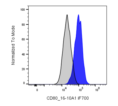
| Recommended Usage | For flow cytometric staining, it is recommended to use less than 0.2 µg of this reagent per 0.5-1.0 million cells in a 100 µL volume. Optimal reagent performance should be determined by titration for each specific application. iF700 has an excitation max at 690 nm and an emission max at 710 nm. |
| Excitation Laser | Red Laser (633 nm) |
| See All Formats | Clone 16-10A1 |
Mouse CD80 (also known as B7-1) is a co-stimulatory molecule expressed primarily on professional antigen-presenting cells (APCs), including dendritic cells, macrophages, and activated B cells. It plays a critical role in initiating and regulating adaptive immune responses by delivering a necessary second signal to T cells during antigen presentation. CD80 expression is upregulated upon immune activation, ensuring that T cell responses are tightly controlled and occur only in the appropriate inflammatory context.
Structurally, CD80 is a type I transmembrane glycoprotein and a member of the immunoglobulin superfamily. It contains two extracellular immunoglobulin-like domains (IgV and IgC), a single transmembrane region, and a short cytoplasmic tail without intrinsic signaling motifs. Despite lacking enzymatic activity, CD80 can influence intracellular signaling indirectly through membrane organization and interactions with other surface proteins on APCs.
The primary ligands for CD80 are CD28 and CTLA-4 (CD152), both expressed on T cells. Binding to CD28 provides a positive co-stimulatory signal that enhances T cell proliferation, cytokine production, and survival. In contrast, interaction with CTLA-4 delivers inhibitory signals that attenuate T cell activation and promote immune tolerance, establishing a balance between activation and suppression.
CD80 is implicated in autoimmune diseases, transplant rejection, and chronic inflammatory conditions when dysregulated. Overexpression can lead to excessive immune activation, while insufficient signaling may impair immune defense.
Therapeutically, CD80 is targeted by agents such as CTLA-4-Ig fusion proteins (e.g., abatacept) that block co-stimulation, providing benefit in autoimmune diseases. Modulating CD80 interactions also plays a key role in cancer immunotherapy and strategies aimed at inducing immune tolerance.
iF700 Armenian Hamster IgG Isotype Ctrl
iF700 Anti-Mouse CD80 Antibody TDS
Have a product or application question? Consult our FAQs or contact us.