| Cat # | Size | Price | Quantity | |
|---|---|---|---|---|
| 515301 | 1 mg | $160 | ||
| 515302 | 5 mg | $400 | ||
| 515303 | 25 mg | $1100 |
| Clone | RB6-8C5 |
|---|---|
| Application | ELISA, WB, Flow cytometry, IHC, ICC, animal model study |
| Host Species | CHO cells |
| Reactivity | Mouse |
| Format | Liquid |
| Target Name | Ly-6G, Lymphocyte antigen 6 complex, locus G, Gr-1 |
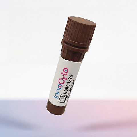
| Product Description | In Vivo Grade Recombinant Anti-mouse Ly6G/Ly6C (Gr-1) Monoclonal Antibody |
| Isotype | Rat IgG2b Kappa |
| Antibody Type | Recombinant |
| Regulatory Status | RUO |
| Purity | >95% by reducing SDS-PAGE |
| Endotoxin | < 1 EU per 1 mg of the protein by the LAL method. |
| Storage Conditions | 4ºC |
| Grade | In vivo |
| Recommended Usage | This product is suitable for in vivo animal use. Optimal amounts need to be determined empirically for each experiment. |
| RRID | AB_3739429 |
| Research Areas | Neutrophils, plasmacytoid Dendritic cells |
| See All Formats | Clone RB6-8C5 |
Ly6G and Ly6C are distinct but closely related cell surface proteins belonging to the Ly-6/uPAR (urokinase-type plasminogen activator receptor) superfamily. In mouse immunology, they are critical differentiation antigens used to identify and classify myeloid cells, particularly neutrophils and monocytes. While often discussed together due to historical antibody cross-reactivity, they mark different cell populations and have distinct biological roles. Structurally, both Ly6G and Ly6C are small, highly glycosylated proteins (approximately 21–25 kDa) that lack a transmembrane domain. Instead, they are tethered to the outer leaflet of the cell membrane via a glycosylphosphatidylinositol (GPI) anchor. This structure allows them to associate laterally with other membrane proteins to transduce signals, despite lacking an intracellular signaling domain of their own.
Unlike classical receptors that bind specific soluble cytokines, Ly6G and Ly6C function primarily through interactions with adhesion molecules, particularly beta-2 integrins (such as CD11b/CD18). They do not have "ligands" in the traditional sense of a hormone or growth factor; rather, they modulate cell adhesion and migration. For example, Ly6G has been shown to regulate the recruitment of neutrophils to sites of inflammation by modulating the function of surface integrins. Similarly, Ly6C is involved in the trafficking of monocytes from the bone marrow to tissues and lymph nodes, influencing their ability to adhere to the endothelium.
In mouse research, these markers are fundamental for distinguishing immune cell subsets, though they are often a source of confusion due to the "Gr-1" antigen. The widely used anti-Gr-1 antibody (clone RB6-8C5) binds to an epitope shared by both Ly6G and Ly6C. Consequently, using anti-Gr-1 depletes or stains both neutrophils (which are Ly6G-high) and monocytes (which express varying levels of Ly6C). To achieve specificity, researchers now use clone 1A8, which binds exclusively to Ly6G, allowing for the specific identification or depletion of neutrophils without affecting monocytes.
Ly6C is particularly valuable for subdividing monocyte populations into functional subsets. "Inflammatory" monocytes are characterized as Ly6C-high (and CCR2-positive), and are rapidly recruited to sites of infection where they differentiate into macrophages or dendritic cells. In contrast, "patrolling" monocytes are Ly6C-low (and CX3CR1-high), functioning to survey the vasculature and promote tissue repair. This dichotomy based on Ly6C expression has become a standard paradigm in studying murine innate immunity and inflammation.
In Vivo Star Anti-Mouse Ly6G/Ly6C (Gr-1) Antibody TDS
Have a product or application question? Consult our FAQs or contact us.