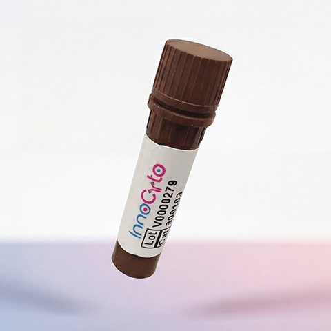
| Cat # | Size | Price | Quantity | |
|---|---|---|---|---|
| 510201 | 1 mg | $160 | ||
| 510202 | 5 mg | $400 | ||
| 510203 | 25 mg | $1100 |
| Clone | 1F4 |
|---|---|
| Application | ELISA, WB, Flow cytometry, IHC, ICC, animal model study |
| Host Species | CHO cells |
| Reactivity | Mouse |
| Format | Liquid |
| Target Name | TIGIT, VSTM3, T-cell immunoreceptor with Ig and ITIM domains, VSIG9 |
| Product Description | In vivo Grade Recombinant Anti-mouse TIGIT Monoclonal Antibody |
| Isotype | Mouse IgG2a Kappa |
| Antibody Type | Recombinant |
| Regulatory Status | RUO |
| Purity | >95% by reducing SDS-PAGE |
| Endotoxin | < 1 EU per 1 mg of the protein by the LAL method. |
| Storage Conditions | 4ºC |
| Grade | In vivo |
| Recommended Usage | This product is suitable for in vivo animal use. Optimal amounts need to be determined empirically for each experiment. |
| Research Areas | Immune Checkpoints, Activated T cells, Tregs, Memory T cells, NK cells, Cancer Immunology, Cancer Marker |
| See All Formats | Clone 1F4 |
T cell immunoreceptor with Ig and ITIM domains (TIGIT) is an inhibitory immune checkpoint receptor that plays a key role in regulating T cell and natural killer (NK) cell responses. TIGIT is expressed on activated CD4+ and CD8+ T cells, regulatory T cells (Tregs), and NK cells. Its primary function is to limit immune activation, maintain peripheral tolerance, and prevent excessive tissue damage during immune responses. In many contexts, TIGIT acts as a brake on cytotoxic and inflammatory activity.
Structurally, TIGIT is a type I transmembrane glycoprotein belonging to the immunoglobulin superfamily. Its extracellular region contains a single IgV-like domain responsible for ligand binding, followed by a transmembrane region and a cytoplasmic tail. The cytoplasmic domain includes an immunoreceptor tyrosine-based inhibitory motif (ITIM) and an immunoglobulin tail tyrosine (ITT)-like motif. Upon ligand engagement and phosphorylation, these motifs recruit intracellular phosphatases and adaptor proteins that mediate inhibitory signaling and dampen activation pathways.
TIGIT binds to several ligands within the poliovirus receptor (PVR) family, most notably CD155 (PVR) and CD112 (PVRL2), which are expressed on antigen-presenting cells, endothelial cells, and many tumor cells. TIGIT competes with the activating receptor CD226 (DNAM-1) for binding to these shared ligands, but binds with higher affinity to CD155. Through both cell-intrinsic inhibitory signaling and competition with co-stimulatory receptors, TIGIT reduces T cell receptor and NK cell–mediated activation, cytokine production, and cytotoxic function.
Dysregulation of TIGIT signaling is implicated in several disease states. In chronic infections and cancer, TIGIT is often upregulated on exhausted T cells and dysfunctional NK cells, contributing to impaired immune control of pathogens or tumors. High TIGIT expression is also a hallmark of suppressive Tregs within the tumor microenvironment, where it reinforces local immune suppression. In contrast, insufficient TIGIT-mediated inhibition may contribute to autoimmune and inflammatory diseases by allowing unchecked immune activation.
Therapeutically, TIGIT has emerged as a promising target in immuno-oncology. Blocking antibodies against TIGIT are being developed to restore effector T cell and NK cell function, particularly in combination with other immune checkpoint inhibitors such as PD-1 or PD-L1 blockade. These combination strategies aim to overcome immune exhaustion and enhance anti-tumor immunity. Conversely, strategies that enhance TIGIT signaling are being considered for autoimmune and inflammatory diseases. Overall, TIGIT represents a key immunoregulatory pathway with significant relevance for both disease pathogenesis and therapeutic intervention.
In Vivo Star Anti-Mouse TIGIT Antibody TDS
In Vivo Star Anti-Mouse TIGIT Antibody, Clone 10A7
Have a product or application question? Consult our FAQs or contact us.