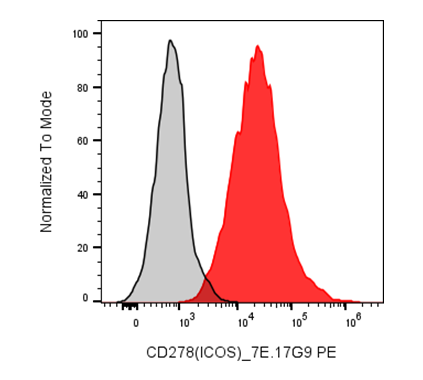
| Cat # | Size | Price | Quantity | |
|---|---|---|---|---|
| 206403 | 25 tests | $55 | ||
| 206404 | 100 tests | $110 |
| Clone | 7E.17G9 |
|---|---|
| Application | Flow Cytometry |
| Reactivity | Mouse |
| Format | PE |
| Target Name | CD278, ICOS, Inducible Costimulatory molecule |
| Isotype | Rat IgG2b |
| Antibody Type | Monoclonal |
| Regulatory Status | RUO |
| Formulation | Phosphate-buffered solution, pH 7.2, containing 0.09% sodium azide and 0.2% (w/v) BSA |
| Protein Concentration | Supplied at a lot-specific concentration. |
| Storage&Handling | The antibody solution should be stored undiluted between 2°C and 8°C, and protected from prolonged exposure to light. Do not freeze. |
| Recommended Usage | For flow cytometric staining, it is recommended to use 5 uL of this reagent per 0.5-1.0 million cells in a 100 µL volume. Optimal reagent performance should be determined by titration for each specific application. PE has an excitation max at 565 nm and an emission max at 575 nm. |
| Excitation Laser | Blue Laser (488 nm) Green/Yellow laser (532/561nm) |
| See All Formats | Clone 7E.17G9 |
Inducible T-cell co-stimulator (ICOS, CD278) is a costimulatory receptor of the CD28 immunoglobulin superfamily that plays a central role in regulating adaptive immune responses. ICOS is expressed at low levels on resting T cells but is rapidly upregulated following T-cell receptor activation on both CD4+ and CD8+ T cells. It is particularly important for the differentiation and maintenance of T follicular helper (Tfh) cells, which provide essential help to B cells during germinal center formation, antibody affinity maturation, and long-lived humoral immunity. ICOS signaling also promotes the function of regulatory T cells (Tregs), Th2 cells, and certain memory T-cell populations while enhancing cell survival, proliferation, and cytokine secretion through activation of the PI3K-AKT signaling pathway.
Structurally, ICOS is a type I transmembrane glycoprotein composed of a single extracellular immunoglobulin variable (IgV)-like domain, a transmembrane region, and a relatively short cytoplasmic tail containing a conserved YMFM motif that recruits phosphoinositide 3-kinase (PI3K). Unlike CD28, ICOS does not possess motifs that strongly induce IL-2 production but instead preferentially supports production of cytokines such as IL-4, IL-10, IL-21, and IFN-γ, depending on the T-cell subset.
The only known physiological ligand for ICOS is ICOS ligand (ICOSL, also known as CD275 or B7-H2), which is expressed on antigen-presenting cells including dendritic cells, B cells, monocytes, macrophages, and some non-hematopoietic cells. Binding of ICOS to ICOSL provides a critical second signal that sustains T-cell activation and promotes effective immune responses.
Dysregulated ICOS signaling has been implicated in autoimmune diseases such as systemic lupus erythematosus, rheumatoid arthritis, and inflammatory bowel disease, where excessive Tfh activity contributes to pathogenic autoantibody production. Conversely, inherited ICOS deficiency results in impaired antibody responses and common variable immunodeficiency. In cancer, ICOS is frequently expressed on activated effector T cells and intratumoral Tregs, making its biological role context dependent. Therapeutically, agonistic ICOS antibodies are being evaluated to enhance antitumor immunity, particularly in combination with PD-1 or CTLA-4 checkpoint inhibitors, whereas antagonistic approaches that block the ICOS-ICOSL pathway are under investigation for treating autoimmune and inflammatory disorders.
PE Anti-Mouse CD278 (ICOS) Antibody TDS
Have a product or application question? Consult our FAQs or contact us.