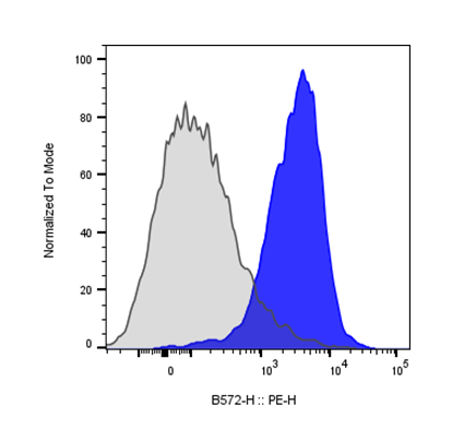
| Cat # | Size | Price | Quantity | |
|---|---|---|---|---|
| 201303 | 25 ug | $55 | ||
| 201304 | 100 ug | $120 |
| Clone | 10F.9G2 |
|---|---|
| Application | Flow Cytometry |
| Reactivity | Mouse |
| Format | Biotin |
| Target Name | CD274, PD-L1, B7-H1 |
| Isotype | Rat IgG2b |
| Antibody Type | Monoclonal |
| Regulatory Status | RUO |
| Formulation | Phosphate-buffered solution, pH 7.2, containing 0.09% sodium azide and 0.2% (w/v) BSA |
| Protein Concentration | 0.2 mg/mL |
| Storage and Handling | The antibody solution should be stored between 2°C and 8°C |
| Recommended Usage | For flow cytometric staining, it is recommended to use less than 0.1 µg of this reagent per 0.5-1.0 million cells in a 100 µL volume. Optimal reagent performance should be determined by titration for each specific application. For detection, use a secondary reagent with this product. |
| RRID | AB_3739038 |
| Research Areas | Activated T cells, Activated B cells, Monocytes, Macrophages, Dendritic cells, NK cells, Immune Checkpoints, Cancer Immunology |
| See All Formats | Clone 10F.9G2 |
Programmed death-ligand 1 (PD-L1), also known as CD274 or B7-H1, is a transmembrane protein that plays a pivotal role in immune regulation by modulating T cell activity. PD-L1 is expressed on a wide range of cells, including antigen-presenting cells, epithelial cells, and many tumor cells. Its primary function is to bind to its receptor, programmed cell death protein 1 (PD-1), located on activated T cells. This interaction delivers an inhibitory signal that reduces T cell proliferation, cytokine production, and cytotoxicity, thereby maintaining immune homeostasis and preventing autoimmunity. However, in pathological contexts such as cancer, PD-L1 expression allows tumor cells to evade immune attack, creating an immunosuppressive microenvironment.
Structurally, PD-L1 is a type I transmembrane glycoprotein belonging to the B7 family of immune checkpoint molecules. The extracellular domain comprises two immunoglobulin-like regions—an IgV-like domain responsible for PD-1 binding and an IgC-like domain that stabilizes the molecule. The protein also contains a single transmembrane helix and a short cytoplasmic tail that lacks classical signaling motifs but may interact with intracellular partners influencing its stability and localization. The PD-L1–PD-1 complex adopts a well-characterized interface where the IgV domains of both molecules interact in a way that blocks T cell receptor-mediated activation signaling.
The main ligands of PD-L1 are PD-1 and CD80 (B7-1). While PD-1 engagement results in T cell inhibition, interaction with CD80 may yield bidirectional signaling effects depending on the cellular context. PD-L1 can be induced by inflammatory cytokines such as interferon-gamma (IFN-γ), linking innate immune responses to immune checkpoint modulation.
PD-L1 plays a major role in numerous diseases. Overexpression of PD-L1 is a hallmark of many cancers, including lung, melanoma, renal, and breast cancers, where it contributes to immune escape. Therapeutically, blocking the PD-1/PD-L1 axis with immune checkpoint inhibitors has revolutionized cancer treatment. Drugs such as pembrolizumab, nivolumab, and atezolizumab disrupt this inhibitory pathway, restoring antitumor T cell function. Moreover, PD-L1 is being explored as both a predictive biomarker for immunotherapy response and a target for novel therapies, including bispecific antibodies and CAR-T cells aimed at enhancing immune-mediated tumor clearance.
Biotin Rat IgG2b Isotype Control Antibody
Biotin Anti-Mouse CD274 (PD-L1) Antibody TDS
Anti-Human PD1 (Nivolumab Biosimilar), Clone Nivolumab
Anti-Human PD1 (Pembrolizumab Biosimilar), Clone Pembrolizumab
Biotin Human PD-L1 (CD274) Protein (C-His-Avi)
Anti-Human PD-L1 (Atezolizumab Biosimilar), Clone Atezolizuma
PE Human PD-L1 (CD274) Protein (C-His)
In Vivo Star Anti-Mouse CD274 (PD-L1) Antibody, Clone 10F.9G2-m2aSL
In Vivo Star Anti-Mouse CD274 (PD-L1) / CD279 (PD1) Bispecific Antibody, Clone 10F.9G2 / RMP1-14
In Vivo Star Anti-Mouse CD370 / CD274 (PD-L1) Bispecific Antibody, Clone 10B4 / 10F.9G2
In Vivo Star Anti-Mouse CD274 (PD-L1) / VEGF-A Bispecific Antibody, Clone 10F.9G2 / B20-4.1.1
In Vivo Star Anti-Mouse CD274 (PD-L1) / VEGF-A Bispecific Antibody, Clone 10F.9G2.1 / G6-23
Have a product or application question? Consult our FAQs or contact us.