| Cat # | Size | Price | Quantity | |
|---|---|---|---|---|
| 514001 | 1 mg | $1750 | ||
| 514002 | 5 mg | $5950 | ||
| 514003 | 25 mg | $17850 |
| Clone | RMP1-14 / B20-4.1.1 |
|---|---|
| Application | Functional assay, Neutralization, animal model study |
| Host Species | CHO cells |
| Reactivity | Mouse |
| Format | Liquid |
| Target Name | PD1, PD-1, PDCD1, CD279, SLEB2, Vascular endothelial growth factor, VEGF-A, VEGFA, VEGF |
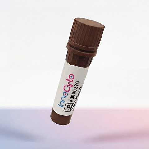
| Product Description | In Vivo Grade Recombinant Anti-mouse PD-1 / VEGF-A Bispecific Antibody |
| Isotype | Mouse IgG2c LALAPG Kappa |
| Antibody Type | Recombinant |
| Regulatory Status | RUO |
| Purity | >95% by reducing SDS-PAGE |
| Endotoxin | < 1 EU per 1 mg of the protein by the LAL method. |
| Storage Conditions | 4ºC |
| Grade | In vivo |
| Recommended Usage | This product is suitable for in vivo animal use. Optimal amounts need to be determined empirically for each experiment. |
| Research Areas | Immune Checkpoints, Activated T cells, Cancer Immunology, Tregs, Tfh cells, Activated B cells |
| See All Formats | Clone RMP1-14 / B20-4.1.1 |
Programmed cell death protein 1 (PD-1) is an immune checkpoint receptor expressed primarily on activated T cells, B cells, and some myeloid cells. It plays a key role in maintaining immune tolerance by downregulating T cell activation when engaged by its ligands PD-L1 or PD-L2 on antigen-presenting cells or tumor cells. Structurally, PD-1 is a type I transmembrane glycoprotein composed of an extracellular immunoglobulin-like domain, a transmembrane region, and an intracellular tail containing immunoreceptor tyrosine-based inhibitory (ITIM) and switch motifs (ITSM). When PD-1 binds to PD-L1 or PD-L2, it initiates inhibitory signaling through SHP-2 phosphatase recruitment, dampening T cell proliferation, cytokine production, and cytotoxic function. In cancer, chronic PD-1 engagement leads to T cell exhaustion, allowing tumor cells to escape immune destruction. This has made PD-1 a central therapeutic target in immuno-oncology, giving rise to checkpoint inhibitors like pembrolizumab and nivolumab.
Vascular Endothelial Growth Factor A (VEGF-A) is a fundamental regulator of angiogenesis, promoting endothelial cell proliferation, migration, and vascular permeability. It binds to tyrosine kinase receptors VEGFR-1 and VEGFR-2 on endothelial cells, driving new blood vessel formation under physiological conditions such as wound healing and tissue repair. In cancer, VEGF-A is frequently overexpressed, leading to aberrant, leaky, and immunosuppressive vasculature that supports tumor growth and hinders immune cell infiltration. By sustaining hypoxic and immunosuppressive microenvironments, VEGF-A contributes not only to tumor progression but also to resistance against immune checkpoint blockade.
A bispecific antibody targeting PD-1 and VEGF-A offers a powerful strategy to simultaneously reinvigorate exhausted T cells and normalize the tumor vasculature. Dual blockade can act synergistically: inhibition of VEGF-A enhances immune cell infiltration by improving vessel structure and reducing hypoxia, while PD-1 blockade restores effector T cell function and anti-tumor immunity. This combination addresses two critical barriers to effective cancer immunotherapy: immune suppression and poor immune access to tumors. Moreover, delivering both mechanisms in a single bispecific antibody may optimize pharmacokinetics, improve co-localization within the tumor microenvironment, and reduce systemic toxicity compared with separate therapies. Preclinical and early clinical studies suggest that PD-1 × VEGF-A bispecifics could yield superior efficacy in “cold” tumors that respond poorly to checkpoint inhibitors alone by converting them into “hot,” inflamed microenvironments more amenable to immune attack. Thus, such bispecifics represent a rational evolution in cancer immunotherapy, bridging vascular and immune modulation for more durable anti-tumor responses.
In Vivo Star Anti-Mouse CD279 (PD1) / VEGF-A Bispecific Antibody TDS
In Vivo Star Anti-Mouse CD279 (PD1) / VEGF-A Bispecific Antibody, Clone 29F.1A12 / B20-4.1.1
Anti-Human VEGF (Aflibercept Biosimilar), Clone Aflibercept
Anti-Human VEGF (Bevacizumab Biosimilar), Clone Bevacizumab
Anti-Human VEGF-A (Brolucizumab Biosimilar), Clone Brolucizumab
Anti-Human PD1 (Camrelizumab Biosimilar), Clone Camrelizumab
Anti-Human PD1 (Cemiplimab Biosimilar), Clone Cemiplimab
Anti-Human PD1 (Nivolumab Biosimilar), Clone Nivolumab
Anti-Human PD1 (Pembrolizumab Biosimilar), Clone Pembrolizumab
Anti-Human VEGF (Fab) (Ranibizumab Biosimilar), Clone Ranibizumab
Anti-Human PD1 (Spartalizumab Biosimilar), Clone Spartalizumab
Have a product or application question? Consult our FAQs or contact us.